-
E-mail
ybiotech@163.com
-
Phone
15026509758
-
Address
Room 310, 3850 Zhoujiazui Road
Shanghai Gubo Industrial Co., Ltd
targetingsystems(targeting systems)DLAR-4
Date: 2021-09-13Read: 2
targetingsystems(targeting systems)DLAR-4
Dual detection of Cypridina Renilla luciferase
Description
Dual luciferase detection reagent based on a single solution:
Save the cost and time of filtering applications
A set of improved ultra sensitive secreted luciferase reporter genes has been developed to analyze the activity of two different promoters in the same group of transfected cells. This method also allows people to study reactions in real-time without killing cells, as these three reporter genes are secreted. Targeting Systems provides a variety of intracellular and secreted dual luciferase detection reagents. The Cypridina Garcia luciferase detection system is particularly attractive because Cypridina luciferase and Gaussia luciferase have strong activity in both intracellular and secretory parts, and both have stable bioluminescence signals when using the reagent for detection. Stability of Renilla luciferase bioluminescence signal (Figure 2).
Cypridina (Vargula) luciferase: Cypridina luciferase (formerly known as Vargula luciferase) from the marine fish Vargula Hilgendorfi is a secreted luciferase with a maximum emission wavelength of 460 nm. It is one of the brightest luciferase enzymes known and has the highest conversion rate.
Gaussia Luciferase: Gaussia Luciferase is a luciferase from the marine copepod Gaussia Princeps (1,2). This ATP free luciferase catalyzes the oxidation of substrate coelicin in light producing reactions and has significant advantages over other luminescent reporter genes, such as secretability and brighter signal intensity, as well as excellent stability of bioluminescence signals. About 10% decays within an hour. The luminescence measured from the supernatant of cultured cells transfected with plasmids expressing GLuc is proportional to the amount of enzyme produced, which in turn reflects the transcription level. Alternatively, cell lysate can be used to measure Gaussia luciferase's high sensitivity even though most of the activity is secreted.
Stability of bioluminescence signals and emission spectra
Figure A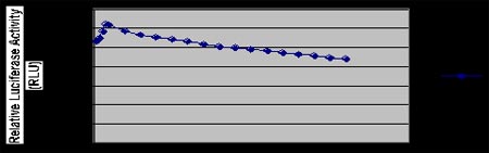
Figure B
DLAR-4 targetingsystems
Figure 2: Dynamics of luciferase activity reported by different luciferase assays using luciferase assay reagents in DLAR-4 system: Establishment of reactions to measure the kinetics of luciferase activity of different luciferase enzymes in transfected cell samples. Measure luciferase activity using DLAR-4 luciferase assay reagent. Figure A: Stability of Cypridina luciferase bioluminescence signal using VLAR component with DLAR-4 dual detection system. Figure B: Stability of GLuc bioluminescence signal using GAR-2 reagent and stabilizer. This reagent is suitable for HTS applications that require the detection of a large number of samples. In the absence of stabilizers, the signal strength is initially slightly higher, but decays faster than with stabilizers. Note: The displayed data is the average of three repeated measurements taken on a Turner TD2020 photometer.
Benefits:
Cypridina Luciferase and Gaussia luciferas both exhibit very strong signals in the supernatant and lysate of transfected cells. Therefore, it can be used as a dual secretory reporter gene or a dual intracellular reporter gene.
Natural Cypridina (Vargula) luciferase and Gaussia luciferase have natural secretion signals and are effectively secreted into cell culture medium after expression. Detecting luciferase does not require cell lysis
Cypridinaia luciferase is one of the brightest luciferase enzymes known, with the highest conversion number and higher bioluminescence signal intensity compared to commonly used firefly luciferase enzymes, making it an ideal transcription reporter gene (1).
The stabilizer component of the Gaussia luciferase detection system provides stable kinetics over a longer period of time, allowing users time for high-throughput analysis and manually delivered testing.
The secreted Cypridina luciferas protein is stable and has * activity in light production, making it highly sensitive for detection (1,2).
Samples containing VLuc and secreted sea kidney luciferase (i.e. transfected growth medium or cell lysate) can be stored for a long time at -20 ° C.
Cypridina-Gaussia Luciferase Dual Assay Reagent-DLAR-4
Package content:
100x Coercetin
Stored at -20 ° C
Gaussia luciferase assay dilution buffer - stored at 4 ° C
Gaussia luciferase assay (GAR) stabilizer - stored at 4 ° C
100x Cypridina Luciferin substrate - stored at -80 ° C
Cypridina substrate dilution buffer - stored at 4 ° C
VLAR buffer (Cypridina luciferase detection buffer) - stored at 4 ° C
Cypridina (Vargula) luciferase detection buffer - stored at 4 ° C
agreement:
Cell supernatant detection:
Gaussia luciferase detection
Dilute 100X colistin to 1X using the required amount of Gaussia luciferase assay dilution buffer (50 ul of dilution reagent is required for each assay).
Transfer 5-20 µ l of sample containing Gaussia luciferase or Cypridina luciferase into each well or photometer tube for detection
Add 8 ul of GAR stabilizer to each sample (this is optional, if the stabilizer is omitted, a higher but less stable signal will be obtained).
Add 50 ul of Gaussia luciferase assay reagent (prepared as described in step 1) to each sample. Mix evenly and read in the photometer.
Cypridina (Vargula) luciferase assay
Dilute 10 µ l of Vargulin substrate to 1 ml using Cypridina (Vargula) substrate dilution buffer.
Transfer 5-20 µ l of sample containing Gaussia luciferase or Cypridina luciferase into each well or photometer tube for detection
Add 40 ul of VLAR detection buffer to each sample.
Prepare 20 ul of diluted Cypridina fluorescein substrate (Vargulin) as described in step 1 and add it to each sample. Mix evenly and read in the photometer.
Determination of luciferase activity in cell lysate: GAR or VLAR luciferase assay reagents can also be used to measure luciferase activity in pre lysed cells. Attention: If you need to measure intracellular luciferase activity, please first lyse the cells using Targeting Systems' cell lysis buffer. (Catalog Number 5X CLR-01)
Dilute 5X CLR buffer with water at a ratio of 1:5.
Suck out the cell culture medium and wash the cells twice with serum-free DMEM.
Add enough 1X cell lysis buffer to cover the cells. Add enough lysis buffer to cover cell.s (50 ul for 96 wells, 300 ul for 12 wells, 800 ul for 6-well culture dishes, and 3 ml for 10 cm culture dishes)
Shake at 400 rpm for 20 minutes on a fixed orbit oscillator (room temperature).
Mix 5-20 µ l of sample or cell lysate containing luciferase with 100 µ l of luciferase assay kit (TS-1) and immediately read in a photometer.
All measuring reagents should be close to room temperature during the measurement.
reference resources:
Yamagishi, K., Enomoto, T., and Ohmiya, Y. (2006) Anal. Biochemistry, 354, 15-21.
Wu, C.、Suzuki-Ogoh, C. 和 Ohmiya, Y. (2007) Biotechniques, 42, 290-292。
Elisa Michelini、Luca Cevenini、Laura Mezzanotte、Danielle Ablamsky、Tara Southworth、Bruce Branchini Spectral analysis gene technology for multiple bioluminescence and high content screening using Aldo Roda * (2007). Anal tightness. Chemistry, 10.1021/ac7016579 S0003-2700 (70) 01657-8
4) BR Branchini, TL Southworth, JP DeAngelis, A Roda, and E Michelini (2006) Fluorescent enzyme from the Italian firefly Luciola italica: molecular cloning and expression. Comp Biochem Physiol B Biochem Mol Biol, October 2006; 145 (2): 159-67.
4) BR Branchini, DM Ablamsky, MH Murtiashaw, L Uzasci, H Fraga, and TL Southworth (2007) developed heat-resistant red and green firefly luciferase mutants for the application of bioluminescence reporter genes. anusstuffyBiochemistry, February 2007: 361 (2): 253-62.