-
E-mail
xue@hkmedqc.com
-
Phone
18148595778
-
Address
111 Shaping North Road, Nanwan Street, Longgang District, Shenzhen
Product Categories
Shenzhen Zhongyukang Technology Co., Ltd


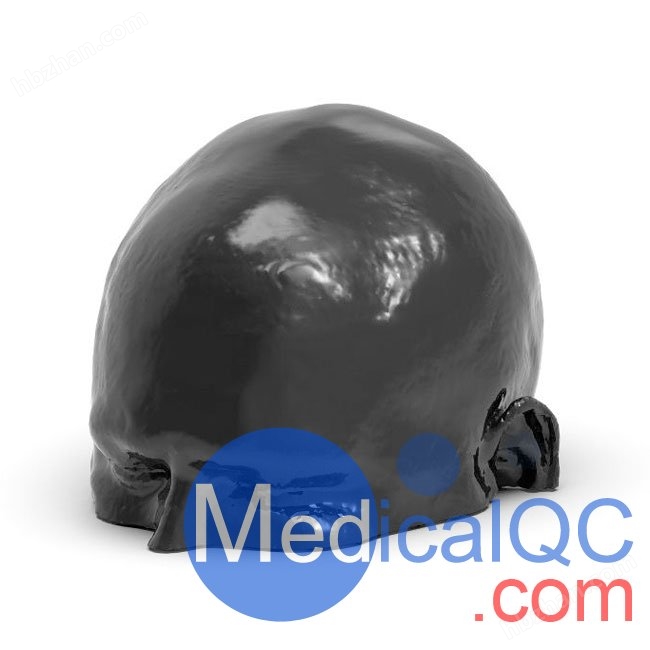
Head phantom of stroke
NegotiableUpdate on 02/06
- Model
- Nature of the Manufacturer
- Producers
- Product Category
- Place of Origin
Overview
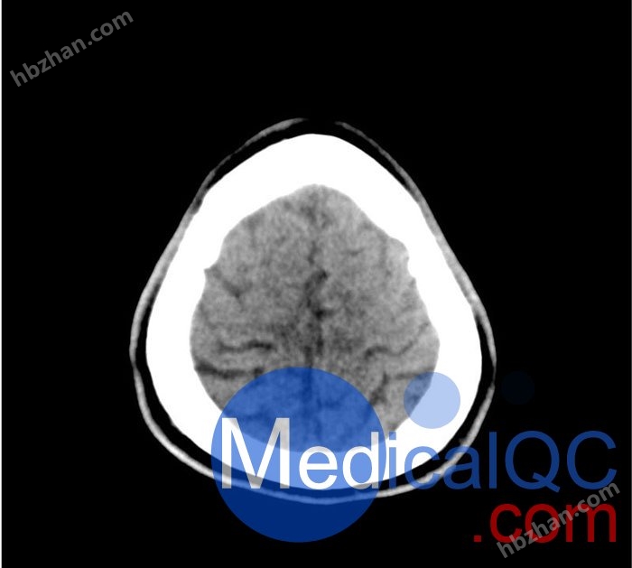
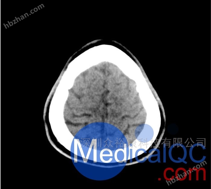
The 50-01 head CT phantom, 50-01 stroke head phantom, and 50-01 cerebral hemorrhage head phantom simulate the head with stroke and hemorrhage patterns. It covers the vertex of the foramen magnum.
Product Details
50-01 Head CT phantom, 50-01Head phantom of strokeThe 50-01 cerebral hemorrhage head phantom simulates a head with stroke and bleeding patterns. It covers the vertex of the foramen magnum.
Stroke patterns include early signs of infarction (high-density middle cerebral artery, disappearance of basal ganglia), acute and subacute watershed infarction, and lacunar infarction at different ages.
The bleeding patterns include subarachnoid hemorrhage, subdural hemorrhage of different ages, intraventricular hemorrhage, and cerebral hemorrhage.
This model can be used for CT (including CBCT) to evaluate and optimize imaging performance and AI supported diagnosis. It is also applicable for training purposes.
This model provides detailed and realistic simulations of common brain pathology, soft tissue, and bone tissue. The air gap is filled with cellulose polymer composite material of about -160HU.
50-01 Head CT Model, 50-01 Stroke Head Model, 50-01 Cerebral Hemorrhage Head Model Diagnostic Features
stroke
Disappearance of high-density middle cerebral artery (MCA) and left basal ganglia
Watershed infarction in the areas of middle/posterior cerebral artery (MCA/PCA) and anterior/middle cerebral artery (ACA/MCA)
8 different age groups of lacunar infarcts
出血
Subarachnoid hemorrhage (2 regions)
Subdural bleeding at different ages (3 regions)
Intraventricular hemorrhage (left ventricle)
Cerebral hemorrhage (2 thalamus, 6 subcortical)
50-01 Head CT Model, 50-01 Stroke Head Model, 50-01 Cerebral Hemorrhage Head Model Specifications
Size: Approximately 190 x 210 x 147 mm
Weight: Approximately 2640 grams
Substrate: Cellulose polymer composite material
Best tube voltage: 120 kVp - adjustable according to requirements
Product applications: organ segmentation, image quality optimization, stroke, intracranial hemorrhage
Diagnostic functions: infarction, bleeding, vascular lesions, spherical lesions
50-01 Head CT Model, 50-01 Stroke Head Model, 50-01 Cerebral Hemorrhage Head Model Imaging Rendering: