-
E-mail
gaoshengkeji@163.com
-
Phone
18128028677
-
Address
101, Building 2, No. 6 Songbailang Xinyuan 1st Road, Dalang Town, Dongguan City, Guangdong Province
Product Categories
Dongguan Gaosheng Electronic Precision Technology Co., Ltd


Ultrasound examination training phantom
NegotiableUpdate on 04/08
- Model
- Nature of the Manufacturer
- Producers
- Product Category
- Place of Origin
Overview
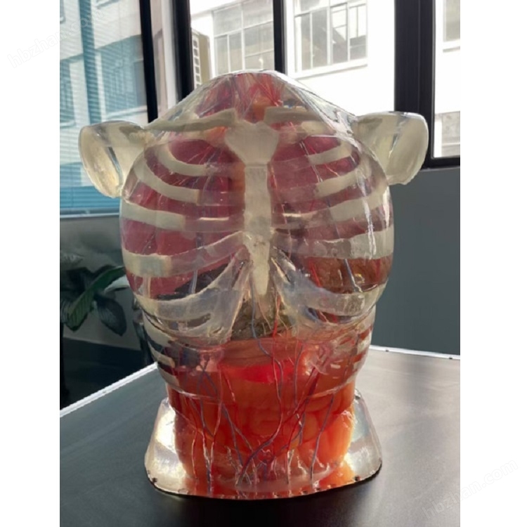
Ultrasound examination training phantom simulates the appearance and structure of the real human abdomen, including the abdominal module from upper to lower abdomen, with surface structures such as bilateral nipples, rib arches, and navel. It has an embedded base shape, a flat base, and meets the requirement of not moving during ultrasound examination. The internal anatomical structure simulation of the phantom includes anatomical structures such as liver, gallbladder, pancreas, spleen, kidney, stomach, abdominal aorta, inferior vena cava, left and right common iliac arteries and veins. The surface should be smooth, without obvious flaws or damage, with the shape and curvature of the abdomen, creating a simulated ultrasound examination scenario.
Product Details
1. Ultrasound examination training phantomProduct Introduction and Structure
1.1 Ultrasound examination training phantomIntroduction
The body simulates the appearance and structure of the real human abdomen, including the abdominal module from the upper abdomen to the lower abdomen, with surface structures such as bilateral nipples, rib arches, and navel. It has an embedded base shape, a flat base, and meets the requirement of not moving during ultrasound examination.
The internal anatomical structure simulation of the phantom includes anatomical structures such as liver, gallbladder, pancreas, spleen, kidney, stomach, abdominal aorta, inferior vena cava, left and right common iliac arteries and veins. The surface should be smooth, without obvious flaws or damage, with the shape and curvature of the abdomen, creating a simulated ultrasound examination scenario.
The anatomical structure of internal organs is clearly visible, allowing for direct observation of their anatomy and location, facilitating the observation of organ morphology and lesion conditions during ultrasound examination.

1.2 Simulation degree
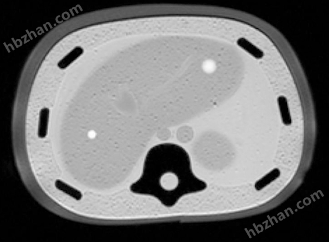
The texture of organs should be close to real human tissue, with appropriate hardness and elasticity. Ultrasonic parameters: sound speed of about 1540-1560m/s, sound attenuation of about 1.45-1.55g/(cm2 · s). Real ultrasound equipment can be used to directly inspect without borrowing software systems, and corresponding tissue ultrasound images can be detected.

Simulate various abdominal organ lesions such as cysts, stones, tumors, etc. within the phantom, which should have different sizes, shapes, and positions to simulate the diversity in real clinical scenarios.

Equipped with a skin color module, it can transparently fit and construct a simulated abdominal appearance, providing simulated abdominal ultrasound examination skill training and obtaining a more realistic operating experience.

1.3 Accuracy
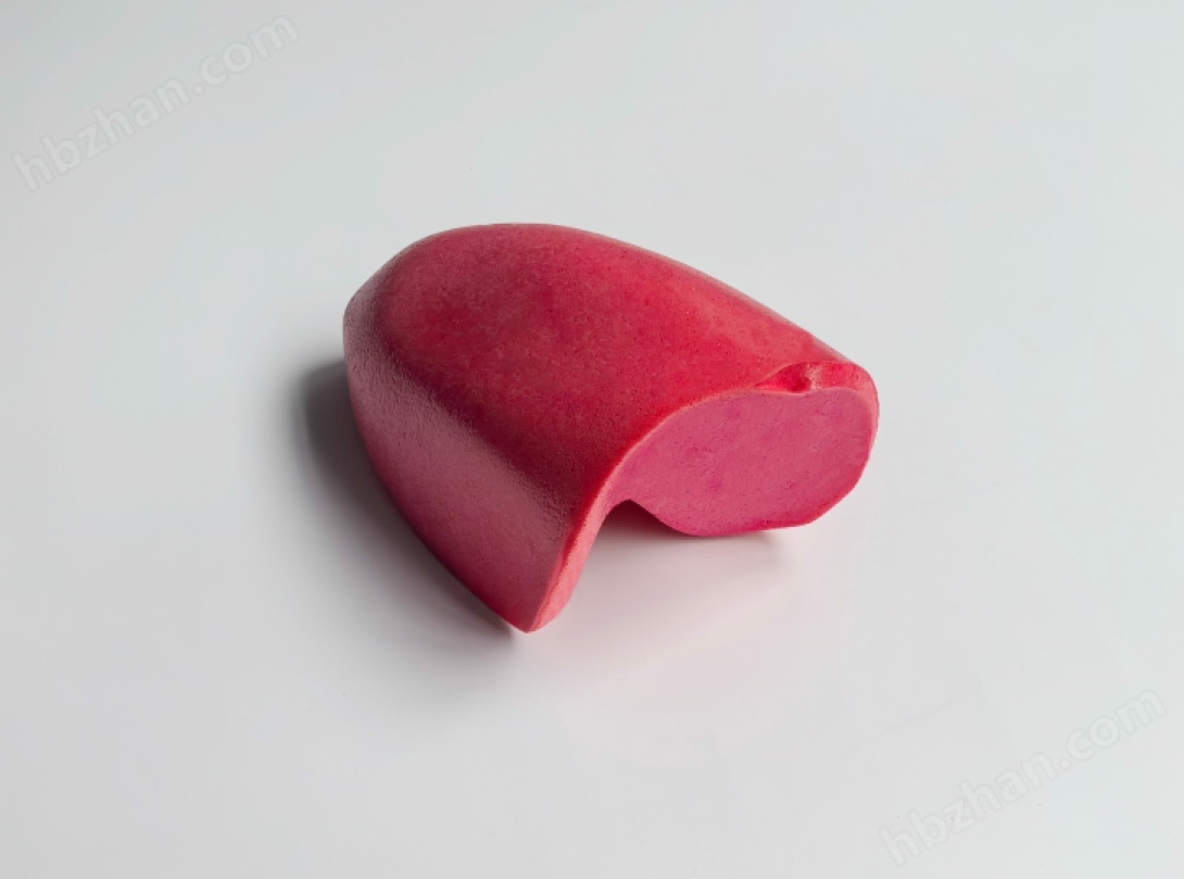
The liver is located in the upper right abdomen and has a left lobe and a right lobe. The important blood vessels that simulate the liver include the portal vein, hepatic vein, inferior vena cava, abdominal aorta, etc. The shape of the blood vessels is similar to that of a real person. Inside the liver, there is a simulated liver tumor and a liver cyst lesion, with the cyst being an anechoic lesion and the tumor being a hyperechoic lesion. During liver ultrasound examination, liver segment exploration and lesion localization training can be performed. The gallbladder recess in the right lobe of the liver contains a gallbladder, simulating cholecystitis and gallstones.

The spleen is located in the upper left abdomen, with a smooth edge and a splenic hilum. Inside the spleen, there is one splenic cyst and one splenic tumor lesion, with the cyst being an anechoic lesion and the tumor being a hyperechoic lesion. The lesion size and location can be explored and measured through the left intercostal space.
The stomach and pancreas are located in the lower abdominal area of the xiphoid process, with the stomach near the abdominal wall and the pancreas behind the stomach. The stomach has a cardia, fundus, body, and pylorus. The pancreas has a uncinate process, head, body, and stomach, as well as pancreatic ducts. There are ≥ 1 pancreatic cyst and ≥ 1 pancreatic tumor lesion, with the cyst being an anechoic lesion and the tumor being a hyperechoic lesion. Gastric and pancreatic ultrasound examination can be performed under the xiphoid process.

The kidneys are located on both sides of the posterior abdomen, with cortex, medulla, and renal pelvis. The cortex has ≥ 1 renal cyst and ≥ 1 renal tumor lesion, while the renal pelvis has ≥ 2 stone lesions. The cysts are hypoechoic lesions, and the tumors are hyperechoic lesions. The bilateral renal hilum is connected to the left and right ureters respectively, and there are stone lesions in the middle of the bilateral ureters. The stones are high echogenicity and can be examined by ultrasound on the outside of the abdomen for kidney and ureteral lesions exploration and description.
It has major blood vessels such as the abdominal aorta and inferior vena cava, with simulated blood inside the vessels. The blood is a hypoechoic fluid dark area, simulating an abdominal aortic aneurysm. Training can be conducted for ultrasound examination of major blood vessels in the abdomen.

1.4 Training Skills
Manual training for abdominal organ ultrasound examination.
Standard sectional examination and lesion identification of abdominal organs.
Abdominal organ ultrasound dissection training.
Assessment of abdominal organ ultrasound examination.
1.5 Operability
The phantom should be easy to install, disassemble, and clean, making it convenient for daily use and maintenance.
The base of the phantom ensures that the phantom remains stable during use.
The ultrasound probe should be able to move smoothly on the surface of the phantom without any obstruction.
1.6 Durability
The phantom should have high durability and be able to withstand repeated use and cleaning.
● The body mold has strong wear resistance and aging resistance to ensure long-term use.

Similar Product Recommend


